Optical Coherence Tomography
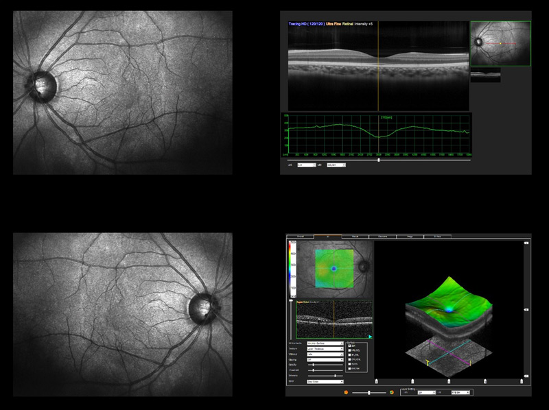
Optical coherence tomography (OCT) is a non-invasive scan of the retina that will provide your doctor at Walter Eye Clinic with detailed, 3D, color-coded, and cross-sectional images of the macula and the retina. These images enable your doctor to detect signs of eye disease in the early stages, which may not have any symptoms.

The OCT scan uses a laser which enables your doctor to obtain high-resolution images of your retina, macula, and optic nerve. You will be asked to sit in front of the OCT machine so that that the scan of your eyes can be completed. Nothing touches your eye, and the test is painless. The test takes about 5–10 minutes to complete.
How are optical coherence tomography (OCT) scans used?
If the doctor at Walter Eye Clinic includes an OCT scan as part of your comprehensive eye exam, this scan can be used to diagnose, manage, or treat several eye diseases and conditions, including the following:
- Diabetic retinopathy
- Age-related macular degeneration
- Macular edema
- Glaucoma
- Drusen
- Retinal detachment, occlusions, or bleeding
- Macular hole
- Neovascularization
If you have a family history or other risk factors for developing eye diseases, the doctor may recommend including an OCT scan with your comprehensive eye exam. If you have already been diagnosed with an eye disease, the OCT scan will provide your Walter Eye Clinic doctor with the information needed to monitor your treatment.
